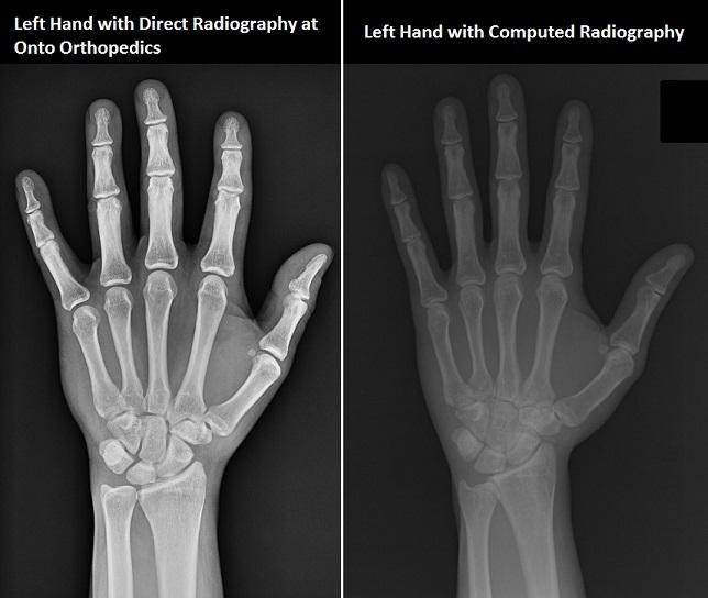
How apparent are these image quality improvements in the final x-ray image? When using the latest generation direct x-ray detector, image quality is usually so much better that even an untrained viewer will notice! Even better, this image quality can often be realized with the same or less dose of radiation when compared with a computed radiography system. Direct radiography systems are also typically much faster- getting you your diagnosis quicker than ever.
You may also ask yourself how these improvements in image quality and dosage could improve the outcome of your treatment. An x-ray typically serves as the first diagnostic test an orthopedic patient receives after an injury. By improving the quality of these images, the orthopedist may be able to forgo further costly and time-consuming tests. Better x-rays may also lead to fewer false-positives.

Many are rightfully concerned about their radiation exposure. Unfortunately though, some confuse CT scans with x-ray scans. Computerized Tomography (or CT) systems take hundreds of x-rays from various angles to build a more comprehensive (and often life-saving!) image of the injured area. In the process however, CT scans can produce tens of times the radiation of a standard x-ray. Rest assured that a standard x-ray image typically results in many times less x-ray exposure than a CT scan. Please see the following link for more: http://www.radiologyinfo.org/en/safety/?pg=sfty_xray In the next article, we will explore more of the differences between our direct radiography x-ray system and a standard CT system.
Related Posts
Cigarettes May Inhibit Inflammation Treatments
Axial spondyloarthritis, also known as AxSpa, is a chronic…


